2013年11月29日 星期五

What a Varicose Vein Looks Like After a Phlebectomy

What a varicose vein looks like

Appears smaller, because the vein contracts and there is no blood flowing through it. This is what a varicose vein looks like after it is removed by phlebectomy. The needle points to the ballooning of the vein (dilatation). This commonly occurs just after the valve, which is not functioning properly. The bulges you see under the skin, in many instances are the dilated areas the needle is pointing to.


Reference information: www.veinexperts.org/
It is not intended as medical advice to any specific person. If you have any need for personal advice or have any questions regarding your health, please consult your doctor for diagnosis and treatment.

2013年11月27日 星期三

How Do Veins Work?

Blood flows through a system of blood vessels, which are veins and arteries. Arteries carry blood rich in oxygen from your heart to all regions of the body. The function of veins is to transport the blood back to the heart. In order for veins to work, the valves must close all the way to prevent backflow of blood. When the muscles in your calf contract; blood is squeezed from the veins. When the muscles in your calf relax, the valves in the veins temporarily close to prevent the blood from flowing away from the heart, in the wrong direction!

In your legs, there are 3 types of veins; superficial, perforating, and deep veins. Think of the perforating veins as connecting the superficial veins to the deep veins. If you are standing, superficial veins run vertically, just beneath the surface of the skin and are often times visible to the naked eye. Superficial veins are responsible for transporting as little as 10% of the blood in the legs. The 10% of blood carried in the superficial veins flow directly into the perforating veins. The perforating veins run perpendicular to superficial veins and again, their function is to connect the superficial veins to the deep veins. Deep veins are located deep within the muscle of your leg and run parallel to superficial veins. Deep veins are responsible for transporting as much as 90% of the blood in your legs back to your heart.

How Veins Work

Reference information: www.veinexperts.org/
It is not intended as medical advice to any specific person. If you have any need for personal advice or have any questions regarding your health, please consult your doctor for diagnosis and treatment.

2013年11月21日 星期四

Varicose Vein Treatment

Common Treatment Options for Varicose Veins

The commonly used treatment options for varicose veins include; compression therapy, thermal ablation, sclerotherapy, phlebectomy and high ligation and stripping. It is important to familiarize yourself with these treatment types and then discuss the best option with your doctor. Most experts in the field of venous disease consider thermal ablation to be the safest, simplest, and most effective treatment type in producing the best long-term results when treating GSV or SSV problems.

Compression Therapy

This conservative treatment is aimed at wearing compression stockings. While this may alleviate some of the symptoms of varicose veins, it does not stop progression of the venous disease. Compression is measured in mmHg. Most people cannot tolerate more than 30 mmHg for prolonged periods of time. Compression therapy is typically used to temporarily relieve symptoms associated with varicose veins and is not a long-term solution to the underlying problem.

Thermal Ablation

This is done with either a laser or radio frequency (RF) catheter. The aim of both of these treatments is it to destroy the vein from the inside using heat. This requires a local anesthetic and an ultrasound. A small needle puncture is made in the saphenous vein. A guide wire is introduced through the needle and over this wire a catheter is placed. The catheter allows for the insertion of the laser filament or (RF) catheter. The filament is directed to just below where the saphenous vein comes off of the femoral vein (3cm below). Next the tumescent solution is placed around the saphenous vein and the laser is then activated and gradually pulled back along the course of the vein. A slightly different technique is used with RF. This heat damages the inside of the vein and over a period of time the vein scars and disappears.
Varicose Vein Treatment Options

Complications are possible with any medical procedure, however, are rare with thermal ablation. There is a very low incidence of deep venous thrombosis (clot) - (DVT) and infections and skin burns are very rare. Most patients have minor issues such as bruising and discomfort in the thigh. Walking soon after a treatment is encouraged. Tumesecent anesthetic is a very dilute solution of xylocaine, sodium bicarbonate, and epinephrine and in most patients, less than 500 cc is needed. A safe dose is over 3 liters.

In this photo, the catheter is placed in the saphenous vein. You can see the laser filament here just before it is placed in the catheter. Once the filament has been placed, tumescent solution is placed around the vein and laser firing begins.


Reference information: www.veinexperts.org/
It is not intended as medical advice to any specific person. If you have any need for personal advice or have any questions regarding your health, please consult your doctor for diagnosis and treatment.

2013年11月20日 星期三

What are Varicose Veins?

Varicose VeinsVaricose veins are large swollen veins that bulge under the skin and are typically blue or green in color. An abnormal valve or a weakened vessel wall causes the bulging. In a normally functioning vein, the valve prevents blood flowing in the wrong direction back towards the foot. In a varicose vein, the valve is damaged and does not close properly. This allows blood to escape backwards when muscles relax. The result is increased pressure in the vein causing it to become swollen with excess blood. Varicose veins can occur anywhere on the leg.
Superficial Vein System 
The Great Saphenous Vein (GSV) and the Small Saphenous Vein (SSV) are the two major veins of the superficial vein system in the legs. The (GSV) is the longest superficial vein which extends from the groin to the foot. The SSV runs from the back of the calf to the lateral ankle. Many superficial veins branch off of both the GSV and the SSV.
If you have a varicose vein, there is a strong probability that it is the result of one of the many valves failing in the GSV/SSV. In a GSV/SSV that functions normally, the valves will close to prevent backflow. When one of the valves functions abnormally in the GSV/SSV, it often causes the GSV/SSV to gradually become larger in diameter. When this happens, the increased pressure causes one of the branching superficial veins that are closer to the surface of the skin to develop into a varicose vein.


 Reference information: www.veinexperts.org/veins/varicose-veins.asp
It is not intended as medical advice to any specific person. If you have any need for personal advice or have any questions regarding your health, please consult your doctor for diagnosis and treatment.

2013年11月15日 星期五

主動脈瘤爆破前有徵狀

步行時突感腹背痛、走路及說話困難甚至頭暈,或是主動脈瘤爆破徵狀。主動脈瘤可出現在胸腔或腹腔,九成個案由血管硬化引起,令動脈如氣球般膨脹,當承受不住血流而爆破,死亡率高達八成。有血管外科醫生指本港每年約有一千宗新症,可透過手術醫治,但港人對該症欠認知,有六成個案到主動脈瘤爆裂才緊急入院,大大減低生還機會。

主動脈是人體最大血管,負責將帶氧血液傳送全身,年齡增長、高血壓、糖尿病、肥胖及吸煙等因素,會令主動脈變得脆弱及硬化,部分血管可膨脹如氣球,形成主動脈瘤,兩至三成屬胸主動脈瘤,其餘為腹主動脈瘤。

美國梅約醫療中心(Mayo Clinic)血管微創中心教授William Stone指,主動脈瘤會令血管壁變薄及失去彈性,一旦無法承受血液流通便會爆破,死亡率達八成。

本港每年千宗新症

香港大學外科學系血管外科主任鄭永強表示,本港每年約有一千宗主動脈瘤新症及一百五十至二百宗死亡個案。隨着港人壽命延長,預計此類血管退化疾病會趨升,而港人飲食西化,糖尿病及高血壓年輕化,可能使主動脈瘤發病年齡下降。

伊利沙伯醫院血管外科副顧問醫生謝卓華指,小部分個案可感到腹部有腫塊「跳跳下」,但多數初時無徵狀,到接近爆破邊緣才感腹背痛及頭暈。本港未如歐美般推行主動脈瘤超聲波普查,因巿民缺乏認識及長者害怕做手術,不少個案均在爆破後才送院。

該院由一九九八至二○○七年共有二百五十三宗病例,急症個案佔六成,立刻做手術仍有三至四成病人死亡,非急症個案的手術死亡率則近乎零。


鄭永強補充,主動脈瘤每年增長約五毫米,當直徑大於五厘米,一年內爆破風險為一成,便應做手術。

醫生會視乎血管瘤形狀及大小,才決定採取開刀或微創手術,前者以人工血管取代血管瘤的動脈,術後需留醫五至九日;微創手術只需在大腿兩側開啟小洞,將支架置入主動脈,以「通波仔」原理令血液流通,只需留院三日,四至六星期可恢復正常活動,但價錢較昂貴,約需九至十萬元。


資料來源:  東方日報
以上所提供的資訊僅作為教育及參考用途,如果你有任何醫療問題,
應向醫生查詢,而不應單倚賴以上提供的資料。

2013年11月12日 星期二

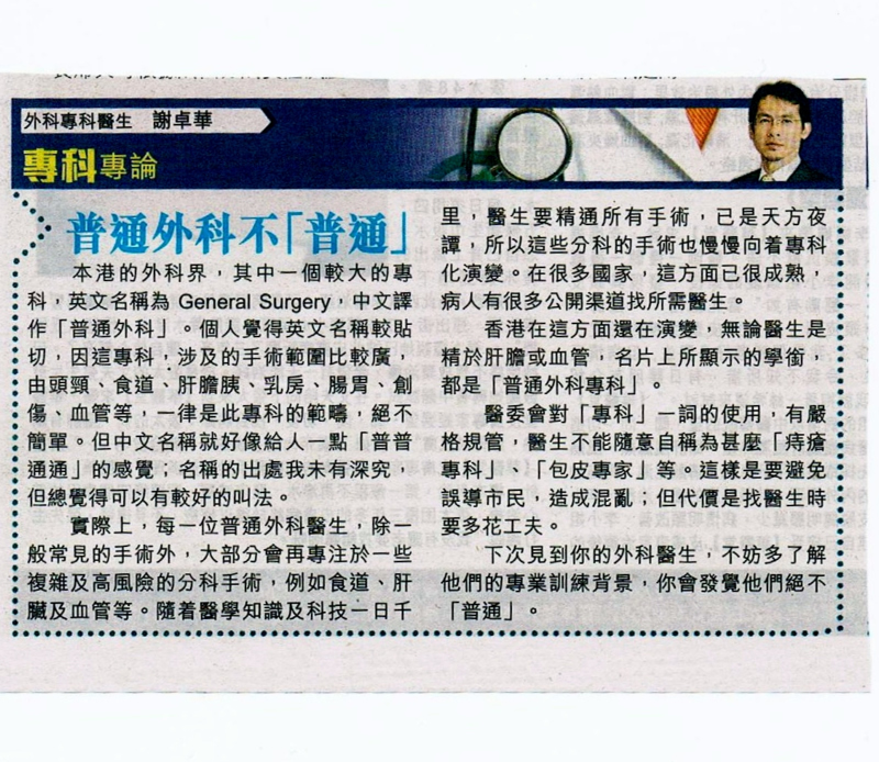
普通外科不「普通」

本港的外科界,其中一個較大的專科,英文名稱為Genereal Surgery, 中文譯作「普通外科」。個人覺得英文名稱較貼切,因這專科,涉及的手術範圍比較廣,由頭頸、食道、肝膽胰、乳房、腸胃、創傷、血管等,一律是此專科的範疇,絕不簡單。但中文名稱就好像給人一點「普普通通」的感覺;名稱的出處我未有深究,但總覺得可以有較好的叫法。

news

實際上,每一位普通外科醫生,除一般常見的手術外,大部份會再專注於一些複雜及高風險的分科手術,例如食道、肝臟及血管等。 隨着醫學知識及科技一日千里, 醫生要精通所有手術,己是天方夜譚,所以這些分科的手術也慢慢向着專科化演變。 在很多國家, 這方面已很成熟,病人有很多公開渠道找所需醫生。

香港在這方面還在演變,無論醫生是精於肝膽或血管,名片上所顯示的學銜,都是「普通外科專科」。


醫委會對「專科」一詞的使用,有嚴格規管,醫生不能隨意自稱為甚麼「痔瘡專科」、「包皮專家」等。這樣是要避免誤導市民,造成混亂;但代價是找醫生時要多花工夫。

下次見到你的外科醫生,不妨多了解他們的專業訓練背景,你會發覺他們絕不「普通」。


外科專科醫生

謝卓華


資料來源:  晴報
以上所提供的資訊僅作為教育及參考用途,如果你有任何醫療問題,
應向醫生查詢,而不應單倚賴以上提供的資料。


2013年11月6日 星期三

How is the diagnosis of Peripheral Arterial Disease?


Peripheral Arterial Disease can be firstly screened by measuring the blood pressure of ankle and brachial, then calculate the Ankle-Brachial Index (ABI). 

If the result of ABI is less than 0.9, it is shown that the case is relatively high chance of suffering from Peripheral Arterial Disease.  For further details, doctors can get more information by performing ultrasound scan or angiography on the affected part.


Reference information: veno.com.hk
It is not intended as medical advice to any specific person. If you have any need for personal advice or have any questions regarding your health, please consult your doctor for diagnosis and treatment.

2013年11月4日 星期一

What problems and sign and symptom do Peripheral Arterial Disease causes?

It is commonly happened in the lower limb arteries. The decreased blood flow due to the narrowed arteries, can lead to the lower limbs pain, numbness, muscle fatigue or cramp. At the early stage of this disease, sign and symptom often occurred during walking, but when stop, the sign and symptom will disappear. This is called “Intermittent Claudication”.

If it is untreated, the blood flow will be nearly or completely blocked due to the plaque is being enlarged by accumulating fat or cholesterol on the inner wall of the affected artery. Limited blood supply will make the surrounding tissue dead, causing gangrene foot, and so increasing the infection risk of the affected limbs. At the late stage, critical ischemic limb can be as serious as amputation is required, in order to saving life under the critical situation. 
                                                                                                      

Those diabetic patients often suffer from Peripheral Arterial Disease. Their nerves are damaged due to diabetes, making patient’s sensation and vision poor. The foot problem is not able to discover easily at the early stage. Ultimately, critical ischemic limb (diabetic foot), one of the Peripheral Arterial Disease also happened on the diabetic patients.


 Reference information: http://veno.com.hk
It is not intended as medical advice to any specific person. If you have any need for personal advice or have any questions regarding your health, please consult your doctor for diagnosis and treatment.